



Presentation
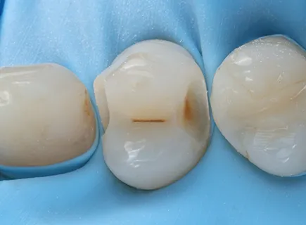
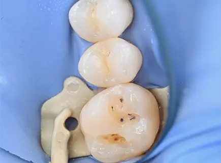
The patient attended for a routine check-up with occasional sensitivity and no visible damage. Clinical inspection found no obvious lesion, but the symptom pattern suggested an early interproximal process.
Diagnostic approach
Transillumination was used to illuminate the tooth from the lingual surface, revealing a shadow consistent with hidden decay. A targeted periapical radiograph confirmed an early carious lesion that had not yet broken through the enamel surface.
This case illustrates why clinical examination alone can miss early interproximal caries — and why transillumination and selective radiographs are part of the diagnostic protocol at MedPalm.
Treatment
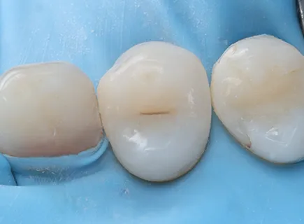
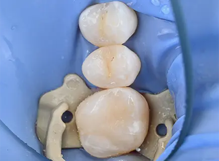
The decayed tissue was removed under rubber dam isolation, which ensures a clean, dry field and the most accurate composite adaptation possible. The cavity was restored with a nanocomposite material matched precisely to the natural tooth shade.
Result
Early detection preserved the majority of healthy enamel and avoided the complications that would have followed if the lesion had progressed: pulp involvement, root canal treatment, and eventually a crown. The patient reported no further sensitivity after treatment.
More Cases
Ready to take the first step?
Book a visit - we'll listen first, then plan your care together

